HOME | DD
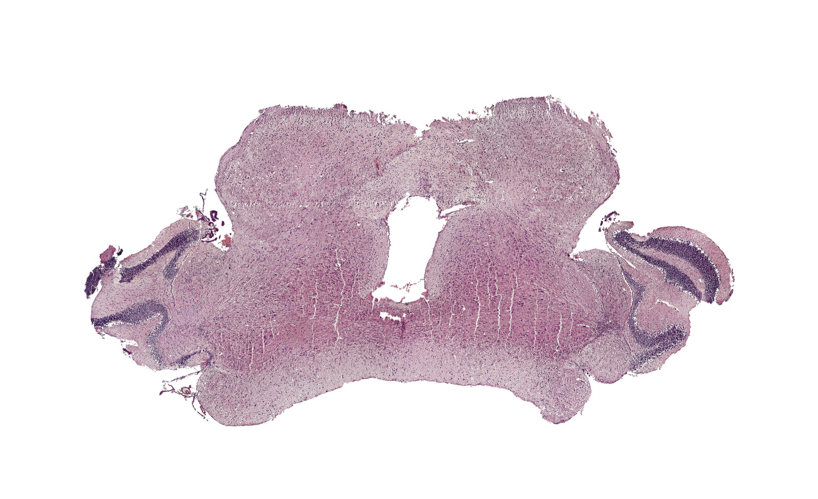
 sumie--dh — Coronal slice of mouse brain
#analysis #anatomy #animal #biology #brain #cell #coronal #histology #laboratory #labrat #method #micrography #microscope #microscopy #mouse #organs #sample #sciart #science #slice #slide #taxidermy #tissue #labwork #purkinje #eosin #cerebelum #scienceart #hematoxylin
Published: 2020-01-29 18:14:48 +0000 UTC; Views: 1449; Favourites: 69; Downloads: 0
Redirect to original
Description
This one is only one which wasn't prepared by me from very beginning. I got this sample at university practice which I attended with desire to correct my possible bad habits and catch few trick from profi histologist. Tissue was already prepared in paraffin block, so I done only fancy job there - slicing on rotary microtome, staining and mounting to Canadian balsam.Pitifully I cannot say much about this one, I don't know age of the mouse nor the strain [but is highly possible that it is B6]. I spent huge time at Mouse Brain Atlas but I'm still unsure about age features of this specimen. Anyway, enjoy nicely visible Purkinje cells between molecular and granular layer in cerebellum - one of largest neurons in brain.
Here is some hint what is visible and which part of brain is on the picture.
cca 45x 125x enlarged , H&E, taken with Carl Zeiss Jenaval.
Bigger and interactive version of this picture is available at Clickable map of this image
Patreon || Ko-fi || Etsy ||
 Twitter || Facebook || Ko-fi || Instagram || Microscopy Discord
 Related content


Comments: 4

I mean daym, it looks like one of those rorschach inkblots.
👍: 0 ⏩: 0

👍: 0 ⏩: 0

👍: 0 ⏩: 0
